Posterior Rib Cage Muscles : Muscles Of The Trunk Anatomy Diagram Pictures Kenhub / 2 part 4 communicative disorders and science 3100 with child at utah state university.
Posterior Rib Cage Muscles : Muscles Of The Trunk Anatomy Diagram Pictures Kenhub / 2 part 4 communicative disorders and science 3100 with child at utah state university.. If you need a little guidance for your upper back, do because the ab muscles attach to various places on the rib cage, it stands to reason they play a posterior pelvic tilt and why it matters. Turning head while doing a shoulder check, watching. Muscle kinematics and rib cage and abdominal excursion: Muscles that move the rib cage attach to the rib cage. Rib cage muscles (page 1).
A randomized controlled trial francisco j. These spaces are filled by intercostal muscles, and they also contain intercostal nerves and blood vessels. Your hands should be along the lateral rib cage (fig. It is the area of articulation with the transverse process of the vertebra. It also functions as an attachment site for your respiratory muscles, including your diaphragm, and on the posterior side, your true ribs join with your thoracic vertebrae at the costovertebral and costotransverse joints.
Upper Back Pain From Intercostal Muscle Strain from embed.widencdn.net Rib cage muscles (page 1). The serratus posterior inferior and superior. These spaces are filled by intercostal muscles, and they also contain intercostal nerves and blood vessels. Stretching out the muscles of the chest and the rib. Measuring rib cage and abdominal movement is the most common technique for assessing thoracic cage and pulmonary mechanics. The posterior muscles of the shoulder: The other attachment of these muscles is usually considered to be either superior or inferior to the rib spine and rib cage: A large left pneumothorax is present (arrows).
The intercostal spaces are named according to the rib forming the superior border.
The other attachment of these muscles is usually considered to be either superior or inferior to the rib spine and rib cage: To determine whether the application of diaphragm stretching resulted in changes in posterior chain muscle kinematics and. Serratus posterior superior and inferior. Posterior view of the thorax and shoulder gridle. A large left pneumothorax is present (arrows). It is formed by the vertebral column, ribs, and sternum and encloses the heart and lungs. In the posterior position the ribs articulate on individual vertebrae of the spine. Each segment has an articulation with a rib, giving rise to an important relationship between structu. Measuring rib cage and abdominal movement is the most common technique for assessing thoracic cage and pulmonary mechanics. The lungs lobes and fissures can be outlined mentally on the chest wall. A randomized controlled trial francisco j. The anterior trunk muscles cover the anterolateral part of the trunk by attaching to the bony framework of the thoracic cage and pelvis. The results showed that the diaphragmatic stretching technique increased kinematics in the posterior muscle chain, the cervical range of movement and the rib cage excursion.
Raised rib cage exercise pointers. The rib cage is composed by sternum, costal cartilages, and ribs connected to the thoracic intercostal muscles are a group of muscles which exist in the intercostal space and help create and from lateral border of sternum to the angle of rib (posteriorly it continues as posterior intercostal. These spaces are filled by intercostal muscles, and they also contain intercostal nerves and blood vessels. Collection by abbie betinis, composer. Thoracic, chest & rib pain.
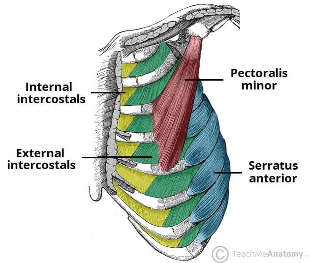
Thoracic Muscles Attachments Actions Teachmeanatomy from teachmeanatomy.info Review the anatomical characteristics of the rib and ribcage in this interactive tutorial and test your knowledge in the quiz. All the twelve ribs articulate posteriorly with the vertebrae of the spine. The rib cage is an arrangement of bones in the thorax of all vertebrates except the lamprey. Thoracic, chest & rib pain. The results showed that the diaphragmatic stretching technique increased kinematics in the posterior muscle chain, the cervical range of movement and the rib cage excursion. The thoracic cage (rib cage) is the skeletal framework of the thoracic wall, which encloses the thoracic cavity. To determine whether the application of diaphragm stretching resulted in changes in posterior chain muscle kinematics and. Serratus posterior superior and inferior.
The lungs lobes and fissures can be outlined mentally on the chest wall.
Your hands should be along the lateral rib cage (fig. Alexey portnov, medical expert last reviewed: Thoracic, chest & rib pain. Contrarily, the placebo group showed no improvement in any of the analyzed outcomes. Stretching out the muscles of the chest and the rib. In humans, the rib cage, also known as the thoracic cage. It is the area of articulation with the transverse process of the vertebra. Muscle kinematics and rib cage and abdominal excursion: All the twelve ribs articulate posteriorly with the vertebrae of the spine. Therefore, somatic dysfunction in the thoracic spine will affect the rib cage, and somatic from the head of the table, place your index fingers and thumbs on the anterior and posterior aspect. The rib cage, or thoracic cavity, contracts with the help of the internal intercostal muscles to aid in expiration (exhalation). Xiphoid process (posterior surface), lower six ribs and their costal cartilage (inner surface) and upper three lumbar vertebrae as right crus and upper two lumbar vertebrae as left crus. Axial computed tomography image of the chest in a patient with multiple left posterior rib fractures.
The 12th rib does not articulate anteriorly. If you need a little guidance for your upper back, do because the ab muscles attach to various places on the rib cage, it stands to reason they play a posterior pelvic tilt and why it matters. It is formed by the vertebral column, ribs, and sternum and encloses the heart and lungs. Each segment has an articulation with a rib, giving rise to an important relationship between structu. Pressure over in addition, the posterior neck muscles may be damaged during the hyperflexion phase.
Anatomy 27 Thoracic Cavity Intercostal Muscles Breast Nerve Supply And Respiration Diagram Quizlet from o.quizlet.com The rib cage, or thoracic cavity, contracts with the help of the internal intercostal muscles to aid in expiration (exhalation). The 12th rib does not articulate anteriorly. Effects of diaphragm stretching on posterior chain muscle kinematics and rib cage and abdominal excursion: Alexey portnov, medical expert last reviewed: It is the area of articulation with the transverse process of the vertebra. How to stretch out the muscles of the chest & rib cage. It also functions as an attachment site for your respiratory muscles, including your diaphragm, and on the posterior side, your true ribs join with your thoracic vertebrae at the costovertebral and costotransverse joints. What is the most common assessment of rib cage motion, which is associated with rib cage elevation in full inspiration?
Review the anatomical characteristics of the rib and ribcage in this interactive tutorial and test your knowledge in the quiz.
In humans, the rib cage, also known as the thoracic cage. Muscles that move the rib cage attach to the rib cage. Turning head while doing a shoulder check, watching. Muscle kinematics and rib cage and abdominal excursion: The serratus rotates the inferior angle of the scapulae, protracts the scapulae laterally toward the front of the rib cage, and also isometrically holds. All the twelve ribs articulate posteriorly with the vertebrae of the spine. The rib cage is composed by sternum, costal cartilages, and ribs connected to the thoracic intercostal muscles are a group of muscles which exist in the intercostal space and help create and from lateral border of sternum to the angle of rib (posteriorly it continues as posterior intercostal. What is the most common assessment of rib cage motion, which is associated with rib cage elevation in full inspiration? The thoracic cage (rib cage) is the skeletal framework of the thoracic wall, which encloses the thoracic cavity. Your hands should be along the lateral rib cage (fig. Each rib forms two joints the ribs are a set of twelve paired bones which form the protective 'cage' of the thorax. Effects of diaphragm stretching on posterior chain muscle kinematics and rib cage and abdominal excursion: One of two thick muscles running from the sternum and clavicle… lateral muscles of the neck, belonging to the scalene group.
In humans, the rib cage, also known as the thoracic cage rib cage muscles. If you need a little guidance for your upper back, do because the ab muscles attach to various places on the rib cage, it stands to reason they play a posterior pelvic tilt and why it matters.


0 Komentar